
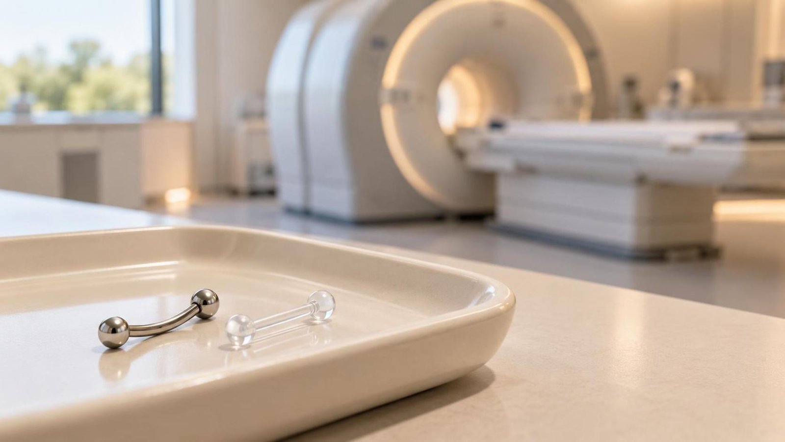
Before an MRI, computed tomography (CT) scan or surgical procedure, anyone with a piercing should tell the healthcare facility and follow the instructions they receive. Whether the jewellery may remain in place or needs to be removed or replaced depends on the procedure, its location, the device and the facility's protocols.
A label such as “implant grade”, a batch certificate, and the fact that jewellery is not attracted to a magnet do not, on their own, establish that a piece is safe for an MRI or may remain in place during a scan or procedure.
Magnetic resonance imaging (MRI)
MRI uses a strong static magnetic field, changing magnetic gradients and radiofrequency pulses. These components present distinct risks: ferromagnetic objects may be attracted to the magnet or displaced; radiofrequency energy may cause heating; and metals may also impair image quality, especially near the area being examined.
Safety therefore cannot be inferred from the alloy name alone. Material, shape, size, position and scan conditions all matter; moreover, a safety classification applies only to the device and conditions actually documented. Ordinary piercing jewellery should not automatically be considered “MR Safe” or “MR Conditional”.
Tell the MRI centre when booking and again during screening, telling staff about every piercing and noting which can be removed and which are recent. Follow staff instructions. Do not use a handheld magnet or preliminary “scout” scan as your own proof that jewellery is safe: assessment is the responsibility of the staff in charge of MRI safety.
CT scans and X-rays
CT scans and X-rays use X-rays, not the strong magnetic field found in an MRI environment. Metal jewellery therefore does not present the MRI-specific risk of magnetic attraction, but it may create artefacts or overlap structures being assessed if it lies within the imaging field. The effect depends on the examination and the jewellery's position.
There is no need to adopt a blanket rule that it “can stay in” or “must always be removed”. Tell the radiologist or radiographer about the piercing and ask whether the jewellery should be removed or changed for that particular examination. Do not change the jewellery without first agreeing how to keep the channel open, if needed.
Surgery
Before surgery, tell the surgical and anaesthetic team about all piercings, including those that are not immediately visible or cannot be removed. The facility may ask you to remove the jewellery for reasons relating to preparation of the operating field, positioning, equipment or the airway. Requirements may vary according to the procedure and local protocol.
During anaesthesia and intubation, oral or nasal jewellery may interfere with manoeuvres or become displaced; the team will assess the risk and give specific instructions. In electrosurgery, it is not accurate to say that every metal piercing will automatically cause a burn: the risk depends on the circumstances and procedure. Do not decide for yourself whether the metal can stay in place; that is for operating-room staff.
Retainers and planning
If the facility asks you to remove jewellery and the channel needs to remain open, contact it in advance to agree whether a retainer is permitted and what type. A retainer is not automatically suitable just because it is non-metallic, flexible or marketed as “medical”: check the material, model, conditions of use and acceptance by the team carrying out the procedure.
- For an MRI, do not insert a retainer or leave one in place without confirmation from the MRI centre. A label or generic statement about the material does not replace assessment of the device and scanning conditions.
- For surgery and anaesthesia, ask the team whether a retainer is permitted and whether it must be removed for any stage of the procedure, for example airway management or preparation of the operating field.
- If the piercing is recent or tends to tighten when the jewellery is removed, consult a qualified piercer in good time, after checking that the facility accepts replacement. The rate of narrowing varies, and it cannot be guaranteed that a channel will stay open for a set period.
- Do not improvise replacements in hospital using cannulas or other devices. Any temporary device or intervention on the channel must be agreed and managed by healthcare staff in accordance with the facility's procedures.
Materials and documentation: what does a certificate show?
ASTM and ISO designations identify material specifications with defined compositions and requirements; examples used for some implant alloys include ASTM F136 / ISO 5832-3 for a titanium alloy and ASTM F138 / ISO 5832-1 for stainless steel. The precise designation is more informative than generic marketing terms such as “surgical steel”.
A Mill Test Certificate (certificate for the starting material) may document the grade and batch of raw material supplied to the manufacturer. On its own, however, it does not necessarily show that an individual finished piece of jewellery came from that batch, nor does it certify that the jewellery is sterile, suitable for everyone, that every aspect of the finished piece complies with the relevant requirements, or that it is safe for a particular MRI scan. Keeping traceability records is useful, but it does not replace clinical screening or the facility's instructions.
In the European Union, Annex XVII of REACH requires nickel release from assemblies intended to be inserted into pierced ears or other pierced parts of the body to be below 0.2 µg/cm² per week. The Commission identifies EN 1811:2023 as the updated reference test method for these assemblies and for articles intended for direct and prolonged skin contact. This compliance concerns nickel release: it is not an MRI safety certification and does not, by itself, authorise use during a CT scan or surgery.
The most useful step is to share the available information about the jewellery with the facility and ask for instructions for the specific examination or procedure. If removal is required, plan any replacement and reinsertion in good time, without prioritising keeping the jewellery in over the healthcare team's instructions.
Sources
- U.S. Food and Drug Administration — MRI: Benefits and Risks.
- National Health Service (NHS) — MRI scan: preparation and screening.
- RadiologyInfo.org — How to Prepare for Your CT Exam.
- Association of Professional Piercers — Jewelry for Initial Piercings.
- Association of Professional Piercers — Preparing for Medical & Dental Procedures.
- Smith F. — Caring for Surgical Patients With Piercings, AORN Journal (2016), abstract in PubMed.
- European Union — Regulation (EC) No 1907/2006 (REACH), Annex XVII, entry 27.
- European Commission — Communication C/2023/1604: EN 1811:2023 standard for nickel release.
Sources consulted on 14 October 2026. This content is for information and does not replace instructions from the imaging centre, surgeon or anaesthetist.